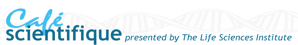
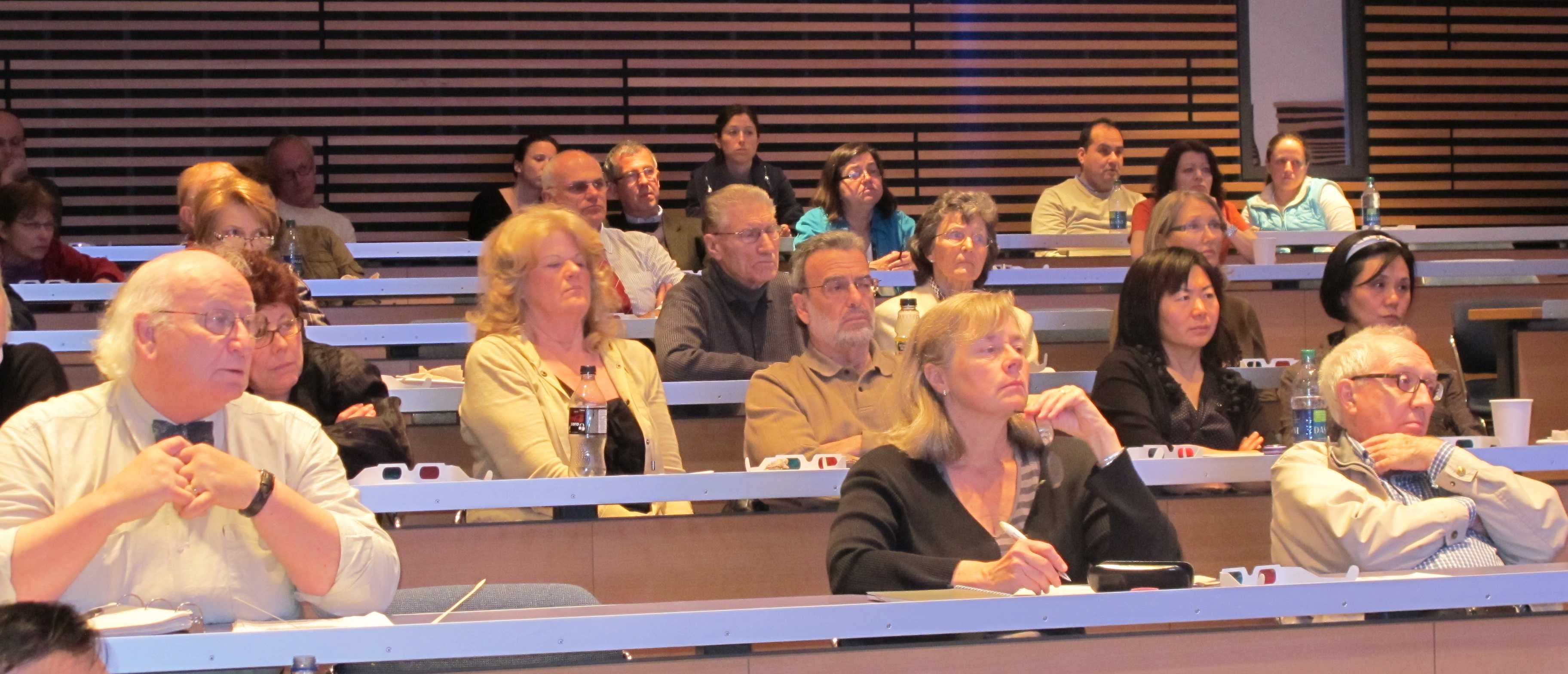
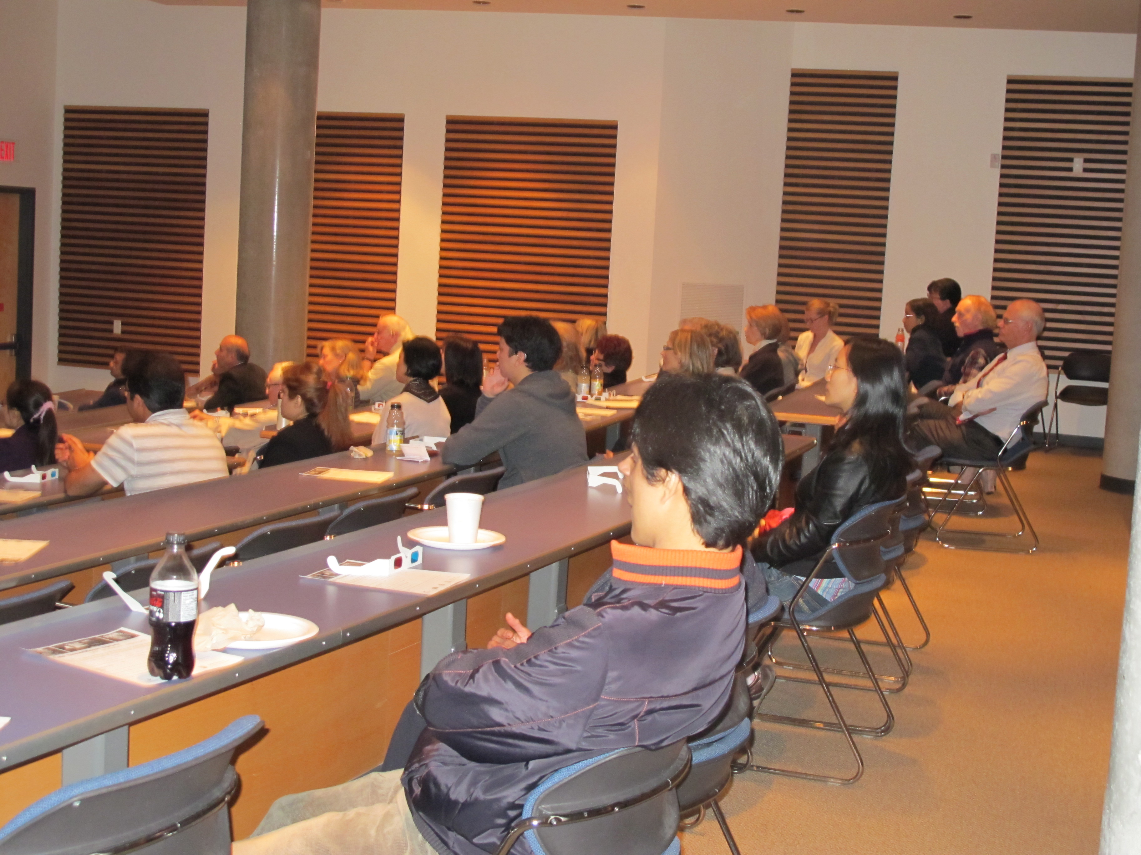
 On May 17, 2011, the Life Sciences Institute (LSI) at the University of British Columbia hosted the seventh LSI Café Scientifique. Over 50 interested community members, as well as students and faculty gathered for an informal and participatory dialogue with experts on the topic of “E-natomy – How digital anatomy has changed the way we teach and practice medicine?”. The Cafes are sponsored by the LSI, The Michael Smith Foundation for Health Research, the Faculties of Medicine and Science and Café Perugia (UBC Food Services).
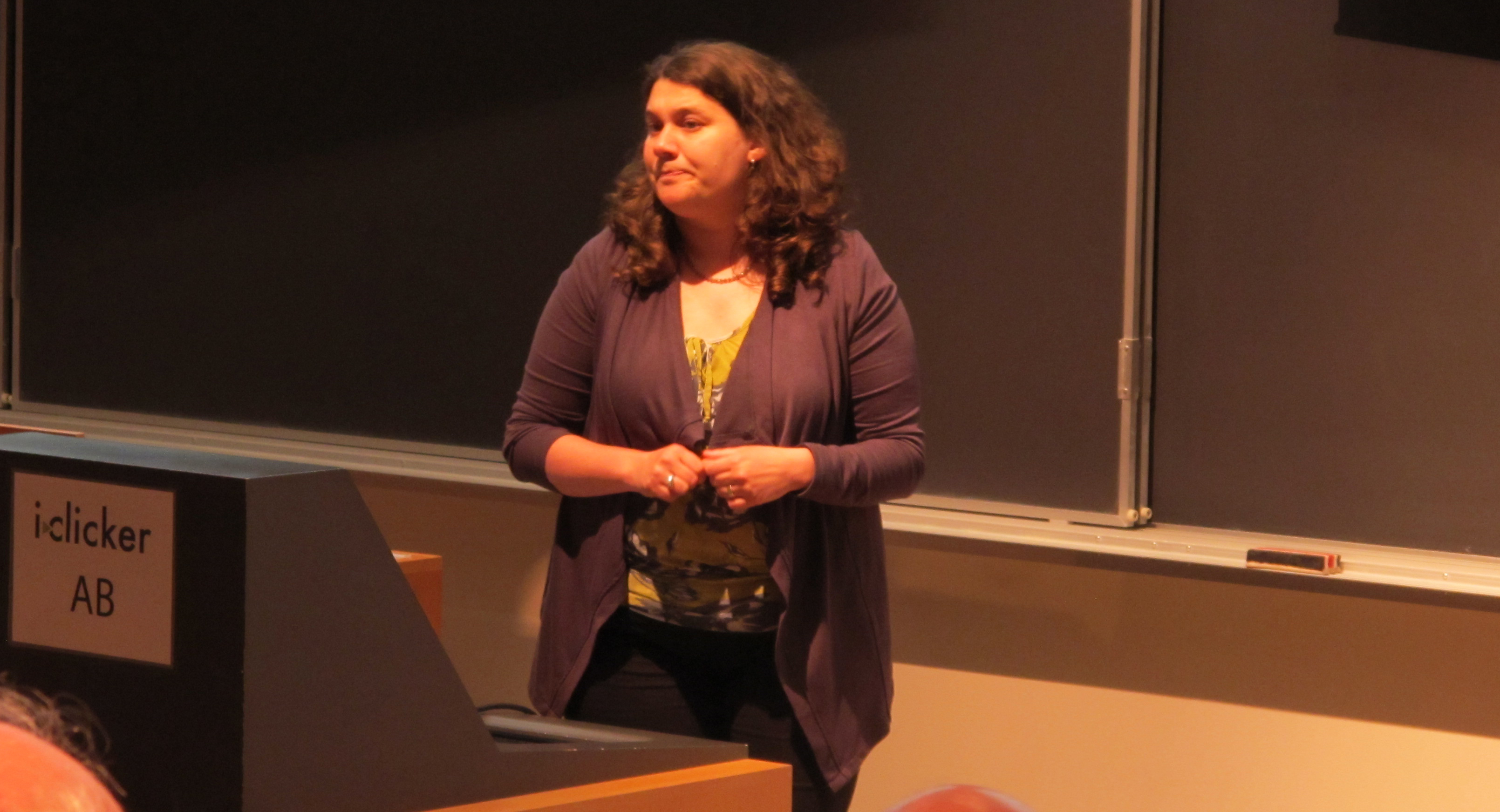
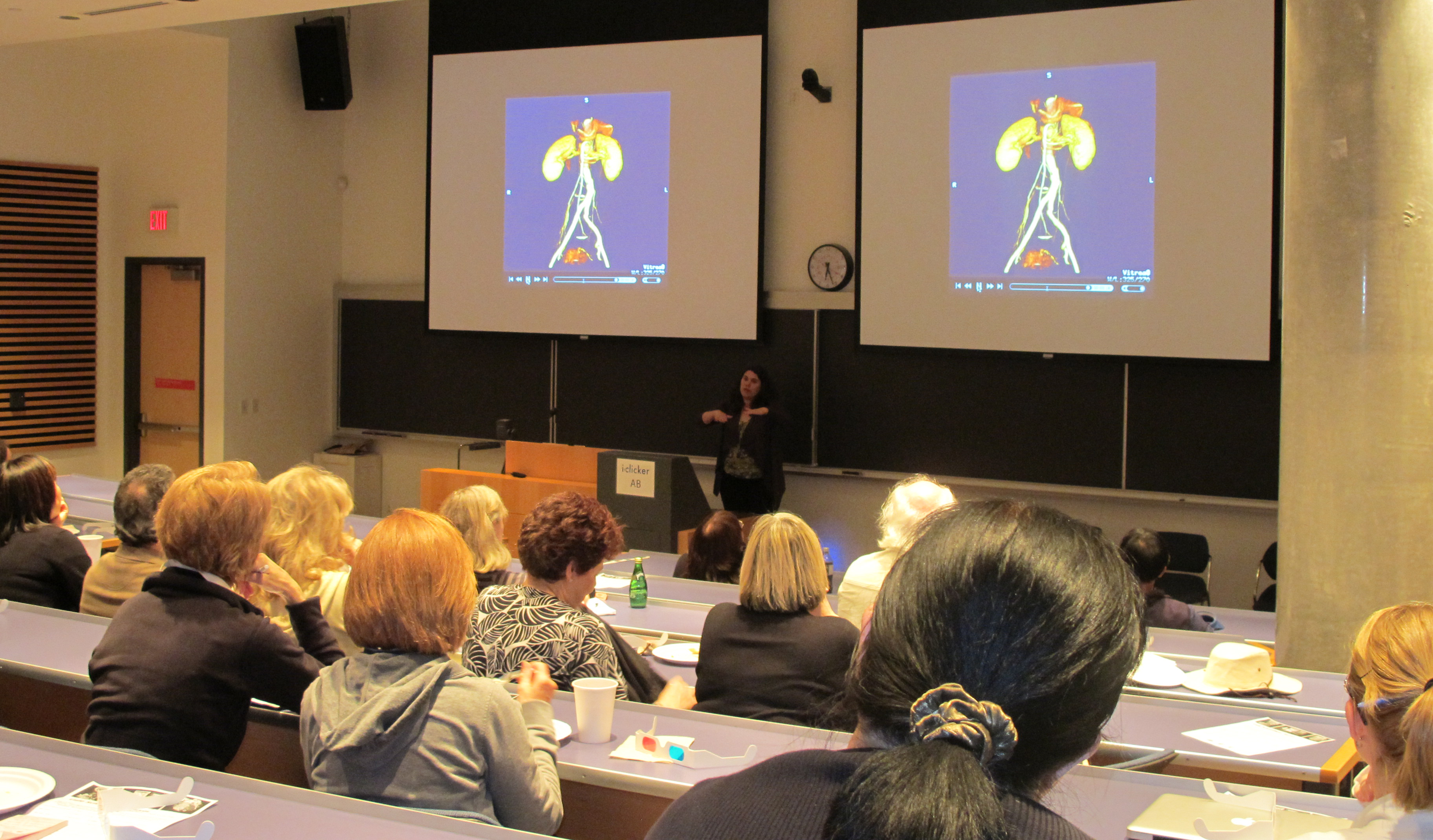
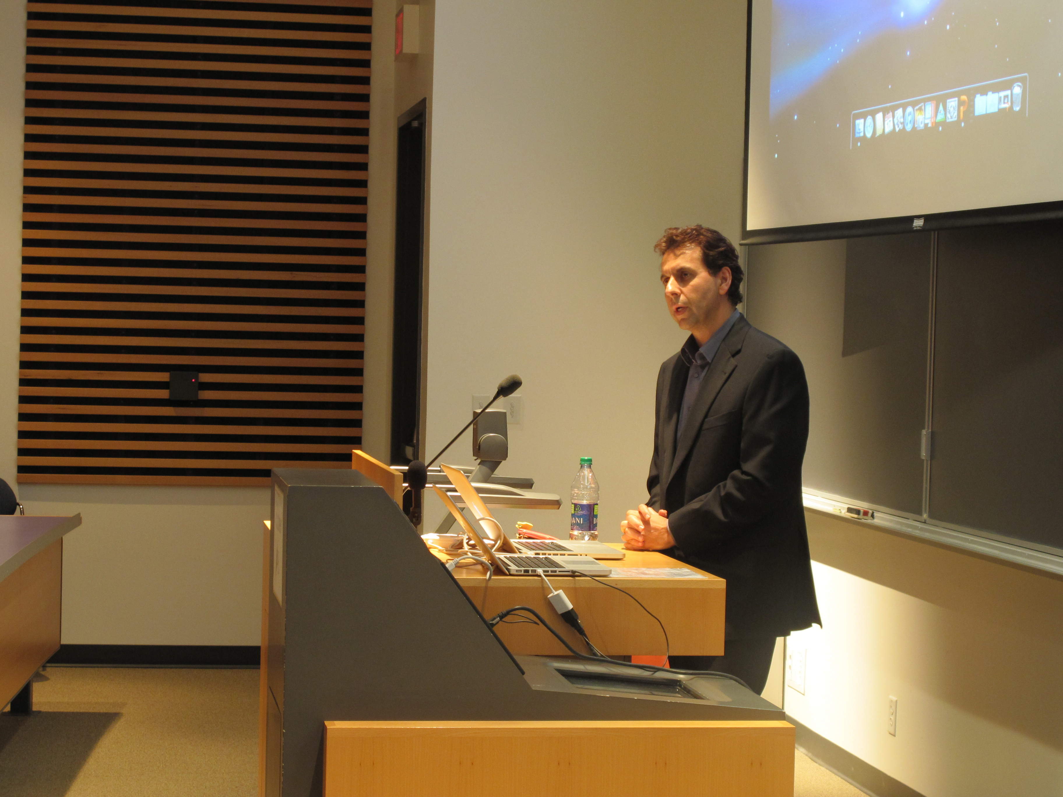
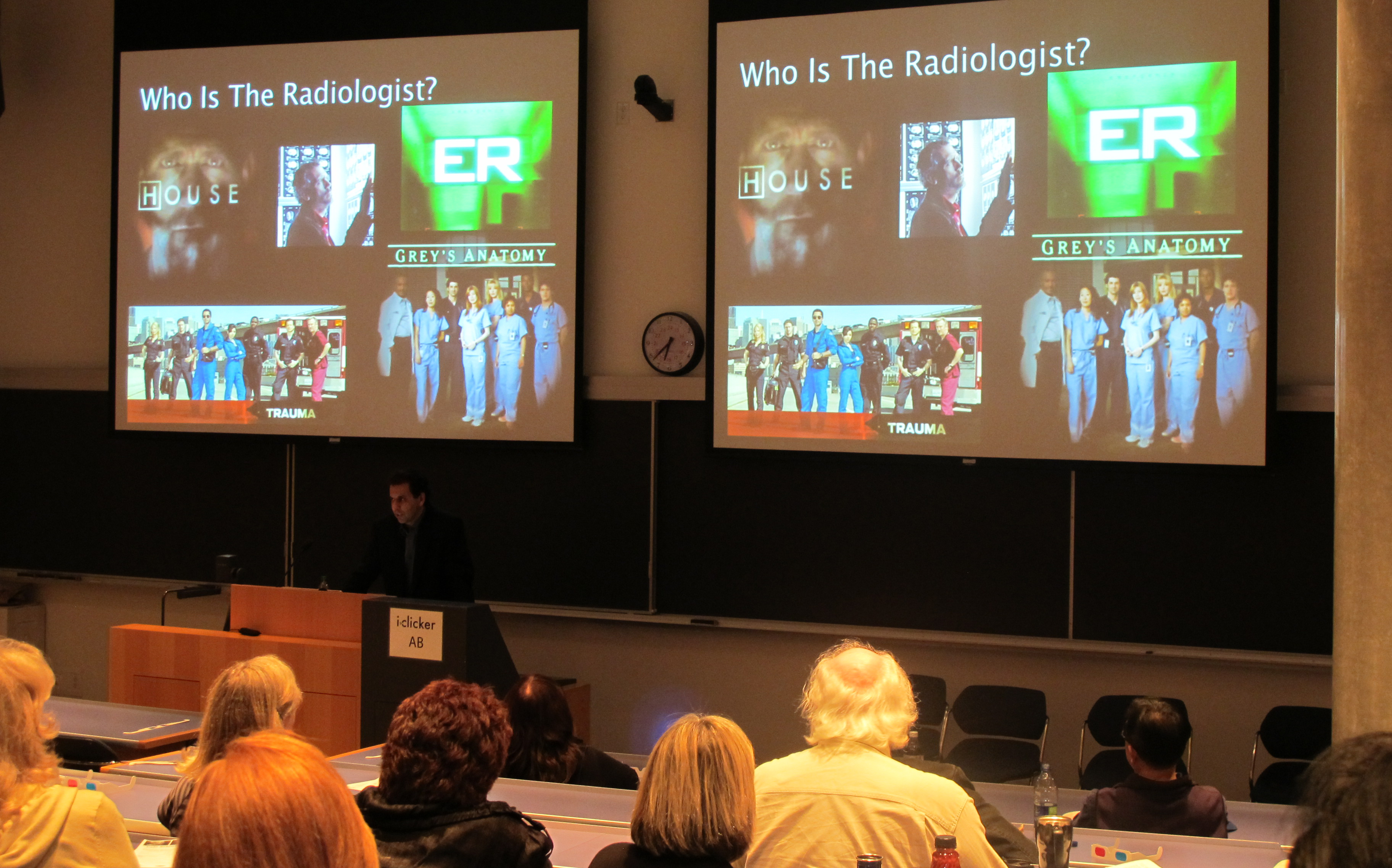
This Café featured two members of the Faculty of Medicine who use digital anatomy in teaching medical students and in clinical practice. This interactive session was presented by Dr. Claudia Krebs, a Senior Instructor in the Department of Cellular & Physiological Sciences, and Dr. Savvas Nicolaou, Associate Professor and Director of Emergency Radiology, practicing at the Vancouver General Hospital. Drs. Krebs and Nicolaou each explained how the advances in whole body imaging have changed the way students learn anatomy and how patients are treated in the ER. Real-life examples of digital anatomy’s role in teaching as well as in practice were shown and the power of CT scans, MRIs and ultrasound scans compared with traditional x-rays was explained.
 New techniques in medical imaging have made it possible to look inside the human body with great precision and with less invasiveness in order to diagnose pathologies that in the past could only be seen with surgical exploration. Learning human anatomy using cadaveric dissection supplemented with x-ray, ultrasound, CT and MRI images allows the physicians of tomorrow to have a better understanding of the 3-dimensional organization of the human musculoskeletal and circulatory systems, internal organs and nervous system. Teaching these new skills in medical school is required to train the modern physician to effectively utilize imaging modalities and interpret pathologies in the clinic today.
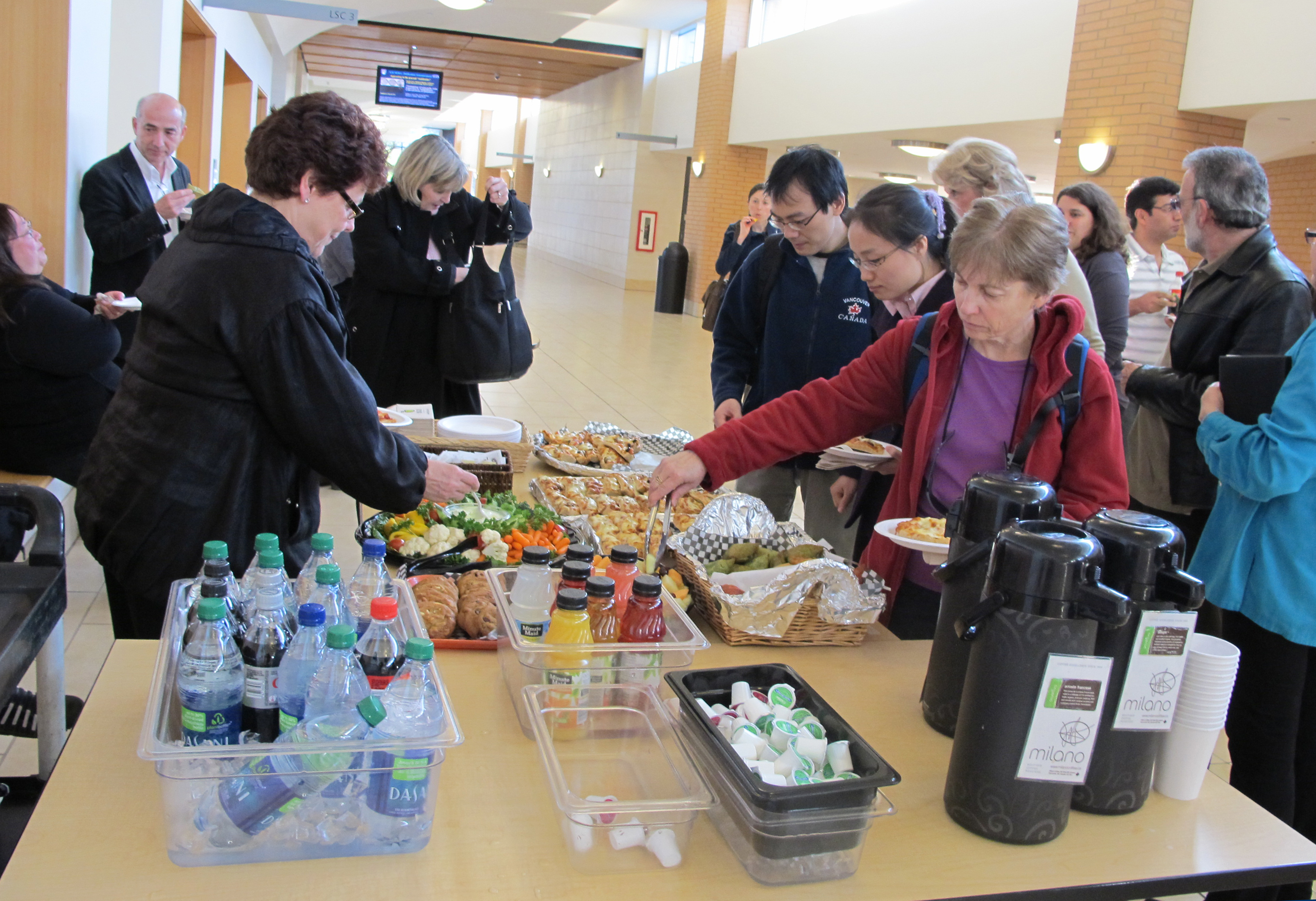
The session was highly interactive and visually entertaining; the presentations included 3D images of the human pelvis, rotating in space which the audience was able to view with 3D glasses. The audience also viewed several staged videos where the power of CT scanning in the Emergency room could be seen as an important diagnostic tool to guide the ER staff in the timely treatment of patients, and in saving lives.
 The door prize awarded to a member of the audience was a copy of the photobook created by Dr. Claudia Krebs with the assistance of Monika Fejtek. This photobook contained digitized images of the original anatomical artwork of the artist Nan Cheney. This collection of pen and ink drawings is an asset of the original Dept of Anatomy at UBC (now called the Dept of Cellular and Physiological Sciences).
Nan Gertrude Lawson Cheney (1897-1985), a contemporary of Emily Carr, was born in Windsor, Nova Scotia and moved to Vancouver in 1937 where she became a well-known portrait and landscape painter. Nan Cheney was also a medical illustrator in the Department of Anatomy at UBC from 1951 to 1956, and continued to work as a medical illustrator until her retirement in 1962. The Department of Cellular and Physiological Sciences currently owns over fifty original works of art by Nan Cheney some of which are exhibited in four glass cases in the main colonnade of the Life Sciences Centre. Each of the works was created from an anatomical dissection done by a staff member in the Department of Anatomy during that time. The Nan Cheney photobook was created to highlight the collection that is displayed in the Colonnade.
